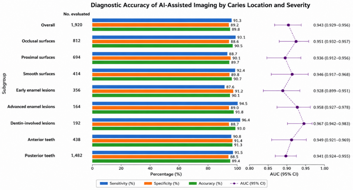
Figure 1: Diagnostic performance of AI-assisted intraoral imaging according to caries location and severity
Asif Rehman1 Mahnoor Khan1*
1Department of Allied Health Sciences, University of Health Sciences, Lahore, Pakistan
*Correspondence: Mahnoor Khan (mahnoorkhanmed@gmail.com)
Received: 20 August, 2026 Revised: 17 September, 2026 Accepted: 19 September, 2026 Published: 30 September, 2026
Background: Early detection of dental caries may prevent lesion progression and facilitate minimally invasive treatment. This study aimed to assess the diagnostic performance of artificial intelligence (AI) supported intraoral imaging in the detection of early dental caries in adults. Methods: This multicenter clinical study included 480 adults from three dental centers. A total of 1,920 tooth surfaces were assessed using AI-assisted intraoral imaging and conventional clinical assessment. The sensitivity, specificity, positive predictive value (PPV), negative predictive value (NPV), accuracy, F1 score, Cohen's κ, and area under the receiver operating characteristic curve (AUC) were evaluated. Results: The reference standard identified 520 (27.1%) surfaces with early dental caries. AI-assisted intraoral imaging showed 91.3% sensitivity (95% CI: 88.6–93.5%), 89.2% specificity (95% CI: 87.5–90.7%), 75.9% PPV, 96.5% NPV, and 89.8% overall accuracy. The AUC was 0.943 (95% CI: 0.929–0.956) and Cohen's κ was 0.747, suggesting good agreement with the reference tests. The sensitivity was highest for lesions involving dentin (96.4%) and was satisfactory for early enamel lesions (87.6%). AI-assisted intraoral imaging had a significantly higher sensitivity (91.3% vs. 76.5%) and AUC (0.943 vs. 0.851; both P<0.001) than conventional clinical evaluation. The mean diagnostic interpretation time was also lower when using AI-assisted intraoral imaging (0.42 ± 0.11 vs. 0.68 ± 0.17 min/surface; P<0.001). Conclusion: AI-assisted intraoral imaging showed high diagnostic accuracy and good agreement with the reference standard for early dental caries detection. Further prospective validation in different populations and clinical settings is warranted before routine widespread use.
Keywords: Artificial Intelligence; Dental Caries; Diagnostic Imaging; Diagnosis, Oral; Machine Learning; Photography, Dental.
Oral diseases, including dental caries, among the most common diseases worldwide and represent a significant burden for people and health care systems 1. Despite being largely preventable, delayed detection may allow the early changes in enamel to become more cavitated and dentinal involvement, pulpal changes and eventual tooth loss 2. The identification of carious lesions is therefore an integral part of the basic tenets of the contemporary minimally invasive dentistry paradigm in which the goal is to be able to identify disease at an early stage where preventive, which aims to identify disease at an early stage when preventive and non-invasive interventions may halt disease progression 3.
Currently, diagnosis of caries is based on a visual-tactile approach, accompanied by a radiographic method where appropriate 4. The diagnostic interpretation might differ based on the clinician's experience, the site of the lesion, image quality and the severity of the disease 5. Limits have led to the advent of ancillary technologies, which can assist in diagnosis and enhance clinical decision-making, and foster greater uniformity of diagnosis 6. Artificial intelligence (AI), specifically machine learning (ML) and deep learning (DL), is a significant innovation in digital dentistry 7. AI systems can analyze a massive number of intraoral images and recognize patterns that are linked to pathological changes that can be hard to detect during routine examination 8. This might consequently enable more objective, reproducible, and rapid evaluation of the surfaces of the teeth and could enhance the efficiency of the identification of early carious lesions, with the support of AI 9. Most importantly, AI should not replace clinical judgment, but rather be used as a tool to assist with diagnosis 10.
Though there has been growing interest in using AI for dental diagnosis, there are uncertainties about its effectiveness in clinical settings. Diagnostic accuracy may vary between institutions, patient populations, tooth surfaces, lesion severities 11. To determine whether promising performance in controlled or single center settings can be sustained across a variety of clinical settings, multicenter clinical evaluation is required. The aim of the present multicenter clinical study is to assess the diagnostic performance of AI-assisted intraoral imaging for the diagnosis of early dental caries in adults. In particular, the study will evaluate sensitivity, specificity, predictive values, overall accuracy, and receiver operating characteristic (ROC) performance, and compared the performance of AI-assisted assessment with conventional clinical assessment.
This Prospective multicenter diagnostic accuracy based study aimed to assess the accuracy of artificial intelligence (AI) supported intraoral imaging in early dental caries detection in adults from the selected dental centers in Lahore and standardized imaging and diagnostic procedures were applied in all centers from September 2024 to September 2025 (ADM/13/2024). The study was conducted according to the principles of diagnostic accuracy research, with AI-assisted intraoral imaging and conventional clinical assessment evaluated as index tests against the same independent reference standard. The study was reported in accordance with the Standards for Reporting Diagnostic Accuracy Studies (STARD) guidelines. A total of 480 adults ≥ 18 years were enrolled, with 160 from each participating center. Inclusion criteria were the presence of at least one natural tooth suitable for intraoral imaging, and the ability to undergo routine dental examination. Patients with a long history of fixed prostheses, significant developmental dental anomalies or images failing predefined image-quality criteria were excluded.
In the study population a total of 1920 tooth surfaces were evaluated. Of those, 520 surfaces were then considered to have early dental caries based on the reference standard. The sample size was calculated using OpenEpi version 3.01 (Emory University, Atlanta, GA, USA; RRID:SCR_021913), assuming 90% sensitivity and specificity, 25% disease prevalence, 95% confidence, and 3% precision. For diagnostic accuracy estimation, the required sample size was calculated using the standard formula n=Z2(1-α/2)Se(1-Se)/(d2P) for sensitivity, where n is the required number of tooth surfaces, Z (1-α/2) is the standard normal deviate corresponding to the confidence level, Se is the anticipated sensitivity, d is the desired absolute precision, and p is the expected disease prevalence. The corresponding specificity-based calculation was also considered, and the larger sample-size requirement was used. After adjusting for within-patient clustering (design effect 1.25), 1,920 tooth surfaces from 480 participants were include 12.
Digital intraoral imaging systems were used to acquire standardized intraoral images in controlled clinical conditions. Images were taken after the removal of obvious debris and saliva contamination if it was needed. To reduce variation due to image acquisition, the same imaging protocol was used at all participating centers. Acquired images were processed using an AI-assisted diagnostic system that detects the visual features related to dental caries. The system produced a classification for each of the tooth surfaces tested (caries-positive or caries-negative). The system also localized and estimated the probability of lesions where applicable. The results of AI were recorded electronically before they were compared to the reference diagnosis.
All participants underwent conventional dental examination by trained dental clinicians. Clinical assessment included visual inspection and tactile examination of tooth surfaces under adequate illumination following routine professional cleaning and drying. Radiographic examination was performed when clinically indicated, particularly where proximal or otherwise clinically uncertain lesions were suspected. Clinical examiners were blinded to the AI-generated classification to minimize incorporation bias. Findings from conventional assessment were recorded independently. Conventional dental examination by trained dental clinicians was done for all participants. The clinical examination was done using visual and tactile methods under adequate illumination after routine professional cleaning and drying of tooth surfaces. Two experienced and calibrated dentists using the International Caries Detection and Assessment System (ICDAS-II) criteria, supplemented by bitewing radiography when clinically indicated. When clinically indicated and when lesions in the proximal areas and/or lesions that were clinically questionable were suspected, the radiographic examination was performed. The incorporation bias was minimized by clinical examiners being blinded to the AI generated classification. Results of traditional assessment were taken independently.
The primary outcome was the diagnostic accuracy of AI-assisted intraoral imaging for detecting early dental caries. Diagnostic performance was evaluated using sensitivity, specificity, positive predictive value (PPV), negative predictive value (NPV), overall diagnostic accuracy, positive and negative likelihood ratios, diagnostic odds ratio, F1 score, Cohen's κ coefficient and area under the receiver operating characteristic curve (AUC). Performance was additionally assessed according to tooth location, surface type, and lesion severity. AI-assisted imaging was evaluated against standard clinical evaluation with the same gold standard. The two approaches to diagnosis were compared in terms of sensitivity, specificity, predictive values, accuracy, AUC, F1 score and agreement statistics. The interpretation time of the diagnostics was also noted with each method. Data analysis was accomplished using IBM SPSS Statistics version 26.0 (Released 2019; IBM Corp., Armonk, NY, USA). Means and standard deviations were used for continuous variables and frequencies and percentages were used for categorical variables. 95% Confidence intervals (CIs) were computed for the diagnostic performance measures.
The discriminative ability of AI-assisted imaging was evaluated using receiver operating characteristic (ROC) curves and the area under the curve (AUC) values was calculated. The agreement between the diagnostic classifications and the reference standard was evaluated using the Cohen's κ statistic. Appropriate paired categorical-data tests were used to compare the difference between the AI assistance and conventional diagnostic performance. A two-sided P value <0.05 was considered statistically significant. All subjects gave written informed consent before starting the study. Analytical datasets were de-identified and images of patients were treated in accordance with institutional policies for data protection and confidentiality.
Overall, AI-assisted intraoral imaging demonstrated high diagnostic performance for the detection of early dental caries across the three participating centers. The baseline demographic and clinical parameters of the 480 adults in the three participating centers on the basis of which the groups were selected are summarized in Table I. The study population was evenly distributed between the centers with the mean age of 32.6 ± 11.4 years and a higher female to male ratio. One thousand nine hundred twenty (1,920) tooth surfaces were assessed, and 520 (27.1%) were considered early dental caries on the basis of the reference standard. Demographic and clinical-history variables were analyzed at the patient level (N=480), whereas tooth-surface characteristics and caries status were analyzed at the surface level (N=1,920).
| Characteristic | Total (N=480) | Center 1 (n=160) | Center 2 (n=160) | Center 3 (n=160) |
| Age, years, mean ± SD | 32.6 ± 11.4 | 31.8 ± 10.9 | 33.2 ± 11.7 | 32.8 ± 11.6 |
| Age 18–29 years | 198 (41.3) | 69 (43.1) | 63 (39.4) | 66 (41.3) |
| Age 30–44 years | 171 (35.6) | 54 (33.8) | 60 (37.5) | 57 (35.6) |
| Age ≥45 years | 111 (23.1) | 37 (23.1) | 37 (23.1) | 37 (23.1) |
| Female | 267 (55.6) | 88 (55.0) | 91 (56.9) | 88 (55.0) |
| Male | 213 (44.4) | 72 (45.0) | 69 (43.1) | 72 (45.0) |
| Teeth/surfaces evaluated | 1,920 | 640 | 640 | 640 |
| Early dental caries identified by reference standard | 520 (27.1) | 171 (26.7) | 177 (27.7) | 172 (26.9) |
| Sound surfaces | 1,400 (72.9) | 469 (73.3) | 463 (72.3) | 468 (73.1) |
| Previous dental restoration | 146 (30.4) | 47 (29.4) | 52 (32.5) | 47 (29.4) |
| Previous dental caries history | 238 (49.6) | 77 (48.1) | 83 (51.9) | 78 (48.8) |
| Regular dental attendance | 291 (60.6) | 99 (61.9) | 94 (58.8) | 98 (61.3) |
Table II shows the overall diagnostic performance of AI supported intraoral imaging for detecting early dental caries. The sensitivity of the AI system was 91.3%, and its specificity was 89.2%, and overall accuracy was 89.8%, with a high negative predictive value of 96.5%. The AUC is 0.943 and Cohen's κ is 0.747, indicating the observed discriminatory performance and agreement with the reference standard, respectively.
| Diagnostic measure | AI-assisted imaging | 95% CI |
| Sensitivity | 91.3% | 88.4–93.7 |
| Specificity | 89.2% | 87.4–90.8 |
| Positive predictive value | 75.9% | 72.4–79.1 |
| Negative predictive value | 96.5% | 95.2–97.6 |
| Overall accuracy | 89.8% | 88.4–91.1 |
| Positive likelihood ratio | 8.45 | 7.15–9.99 |
| Negative likelihood ratio | 0.10 | 0.07–0.13 |
| Diagnostic odds ratio | 84.5 | 58.2–122.7 |
| Area under ROC curve | 0.943 | 0.929–0.956 |
| F1 score | 0.828 | 0.807–0.848 |
| Cohen's κ | 0.747 | 0.716–0.778 |
The diagnostic accuracy of AI-supported imaging by caries location and caries severity is shown in Table III. Surface-location analyses were performed for occlusal, proximal, and smooth surfaces, whereas tooth-location analyses were performed separately for anterior and posterior teeth. Lesion-severity categories were assigned according to the predefined reference-standard criteria. Dentin-involved lesions had the highest sensitivity (96.4%) and the sensitivity for early enamel lesions was comparatively lower (87.6%) which indicates that lesions with subtle early changes are more difficult to diagnose
| Subgroup | No. evaluated | Sensitivity (%) | Specificity (%) | Accuracy (%) | AUC (95% CI) |
| Overall | 1,920 | 91.3 | 89.2 | 89.8 | 0.943 (0.929–0.956) |
| Occlusal surfaces | 812 | 93.1 | 88.6 | 90.5 | 0.951 (0.932–0.967) |
| Proximal surfaces | 694 | 88.7 | 90.1 | 89.7 | 0.936 (0.912–0.956) |
| Smooth surfaces | 414 | 92.4 | 89.8 | 90.7 | 0.946 (0.917–0.968) |
| Early enamel lesions | 356 | 87.6 | 91.2 | 90.1 | 0.928 (0.899–0.951) |
| Advanced enamel lesions | 164 | 94.5 | 89.0 | 91.8 | 0.958 (0.927–0.978) |
| Dentin-involved lesions | 192 | 96.4 | 88.7 | 93.0 | 0.967 (0.942–0.983) |
| Anterior teeth | 438 | 90.8 | 91.4 | 91.3 | 0.949 (0.921–0.969) |
| Posterior teeth | 1,482 | 91.5 | 88.5 | 89.4 | 0.941 (0.924–0.955) |
Figure 1 illustrates the diagnostic performance of AI-assisted imaging across different caries locations and severity levels, demonstrating consistently high sensitivity, specificity, accuracy, and AUC values across all subgroups.
Figure 1: Diagnostic performance of AI-assisted intraoral imaging according to caries location and severity
Table IV compares the intraoral imaging by AI to the conventional clinical examination. AI was significantly more sensitive (91.3% vs. 76.5%), more accurate as measured by AUC (0.943 vs. 0.851), and had a higher negative predictive value than conventional assessment. Additionally, the AI-assisted assessment also involved less time interpreting the results, indicating potential benefits for earlier detection of lesions and for streamlined clinical decision making.
| Diagnostic outcome | AI-assisted imaging | Conventional clinical assessment | P value |
| Sensitivity | 91.3% | 76.5% | <0.001 |
| Specificity | 89.2% | 92.1% | 0.041 |
| Positive predictive value | 75.9% | 78.4% | 0.284 |
| Negative predictive value | 96.5% | 90.3% | <0.001 |
| Overall accuracy | 89.8% | 87.9% | 0.018 |
| AUC | 0.943 | 0.851 | <0.001 |
| F1 score | 0.828 | 0.773 | <0.001 |
| Cohen's κ | 0.747 | 0.681 | 0.006 |
| Missed early caries lesions, n (%) | 45 (8.7) | 122 (23.5) | <0.001 |
| False-positive classifications, n (%) | 151 (10.8) | 110 (7.9) | 0.031 |
| Mean diagnostic interpretation time, min/surface | 0.42 ± 0.11 | 0.68 ± 0.17 | <0.001 |
The AUC for AI-assisted imaging was computed from the continuous AI-generated probability scores while the AUC for conventional clinical assessment was computed from the ordinal ICDAS-II scores recorded during clinical examination. The differences between the two AUCs were evaluated with a patient-level clustered bootstrap procedure, with the subjects resampled as clusters to take into account the correlation between multiple tooth surfaces from a single subject. All comparisons were made on the same tooth surfaces with the same independent reference standard. Surface-location analyses were done in each of the following locations separately: occlusal, proximal, and smooth. Tooth-location analyses were done in the anterior and posterior. The lesion-severity categories were assigned based on the pre-established ICDAS-II criteria.
This multi-center study showed the high diagnostic performance of AI-assisted intraoral imaging for early dental caries diagnosis, with a sensitivity of 91.3%, specificity of 89.2%, accuracy of 89.8% and AUC of 0.943. Additionally, the high NPV (96.5%) and good agreement with the reference standard (κ=0.747) support good caries-positive surface exclusion capacity. Overall, these results indicate that AI-aided image interpretation could enhance the detection of subtle lesions that are difficult to identify when visual inspection is performed in a routine manner. Meanwhile, the discrimination obtained using standardized intraoral photographs was also high at 0.964, with sensitivity of 89.6% and specificity of 94.3% 13. Similarly, Kang et al. reported excellent discrimination of 95.2% AUROC and 89.3% specificity using intraoral-camera images with an ensemble Inception-ResNet-v2 model 14.
The PPV value of 75.9% may be due to a few AI-positive surfaces being identified in sound surfaces, while the NPV value of 96.5% suggests that the negative classifications were reasonably accurate in this study population. However, PPV and NPV are influenced by disease prevalence; therefore, these values may vary when the prevalence of dental caries differs across populations. Recent reviews highlighted large variability due to differences in the imaging devices, annotation methods, lesion thresholds, and datasets and outcome measures 15. A study reported that there are significant differences in F1 score between various professional and intraoral cameras and smartphone cameras 16. The lower sensitivity found for early enamel lesions (87.6%) as compared to dentin-involved lesions (96.4%) is clinically important as the image characteristics produced by early enamel lesions are subtler. The feasibility of deep learning classification of caries from intraoral photographs using a smartphone; other imaging modalities also showed variations with different stages of caries 17. A study demonstrated a deep-learning system on bitewings with a sensitivity of 0.89 for proximal lesions in deeper dentin lesions 18. The present findings thus indicate that further modelling should focus specifically on the recognition of early noncavitated enamel changes.
This is linked to how AI could be a valuable supplementary tool for clinical diagnostics, as is evident in the comparisons with traditional assessment methods 19. The present findings therefore support the use of AI as an adjunctive diagnostic tool in clinical practice. The present study results indicate that AI is a tool for the dentists to assist in their diagnosis, which means that AI can be used as a decision support tool but it cannot take the place of clinical experience and judgment 20. However, as the image acquisition method, the threshold of lesions and the type of model differ, it is difficult to compare the results of the present study with the other studies. Differences in reference standards and study populations may also contribute to variation between studies.
There are a number of limitations. The multicenter design enhances applicability, but the image sample was limited to three dental centers and due to the exclusion of poor-quality images and some complex dental conditions, results may not be generalizable 21. Additionally, no assessment was conducted to see if AI-assisted diagnosis would alter treatment plans or outcomes. Future prospective studies should include larger and more diverse populations, independent external validation, standardized lesion definitions and thresholds across imaging devices, and formal assessment of clinician–AI interaction and patient-level outcomes
The sensitivity and specificity of AI-supported intraoral imaging were good for detecting early dental caries and there was significant agreement with the reference standard. It also was more sensitive and faster to interpret than conventional clinical evaluation. The results suggest that AI-assisted intraoral imaging could be a valuable tool to enhance the diagnosis and efficiency of early caries detection. These results need to be confirmed in larger, more diverse groups of patients and through external validation in different clinical settings prior to routine use.

Journal of Dentistry and Oral Sciences Research, J Dent Oral Sci Res 2026:1(1), p10-14 (jdosr.org) © 2026 Authors. This work is published by Multidisciplinary Scholarly Advancement and Research MSAR Institute. The full terms of Journal Publishing policy is available at https://jdosr.org/index.php/jdosr/publishing-policy and incorporate the Creative Commons Attribution – Non Commercial (CC BY, NC 4.0) License https://creativecommons.org/licenses/by-nc/4.0/. By accessing the work you hereby accept the Terms. Non-commercial uses of the work are permitted without any further permission, provided the work is properly attributed. Publisher’s Note: MSAR Institute remains neutral with regard to jurisdictional claims in published maps and institutional affiliations, and assumes no liability for the scientific accuracy or clinical efficacy of the content herein, as they rest entirely with the authors.