ORIGINAL ARTICLE
Comparative Effect of Minimally Invasive Endodontic Access Cavity Designs on Fracture Resistance and Root Canal Treatment Outcomes
Naila Anjum1* Inam Ur Rehman2 Faiza Ikram3
1Department of Nursing, Farooq Hospital Lahore, Pakistan | 2Dental Sciences, Superior University, Hospital Lahore, Pakistan | 3Department of Medicine, Down University of Health Sciences / Private Dental Assistant, United Kingdom
*Correspondence: Naila Anjum (nailaanjum951@gmail.com)
Acknowledgement: None;
Competing Interests: The author declared no competing interests;
Grant Support and Funding Source: No external funding source;
Study Ethical Approval: Institutional Head approval was obtained as per authors’ declaration before the conduct of the study (MS-014/EC2022);
Consent for Participation and Publication: Written informed consent was obtained from participants;
Availability of Data and Materials: Available upon reasonable request from correspondence author;
Use of Artificial Intelligence: The corresponding author declared that no artificial intelligence or AI-assisted tools were used in any stage of manuscript preparation and editorial processing;
Authors’ Contribution: NA: Conception, study design, literature search, critical review, drafting the manuscript, approval of the final version to be published. IUR, FI: Data acquisition, statistical analysis, data interpretation, critical review, drafting the manuscript, approval of the final version to be published. All Authors agree to be accountable for all aspects of the work in ensuring that questions related to the accuracy or integrity of any part of the work are appropriately investigated and resolved.
Received: 25 August, 2026 Revised: 17 September, 2026 Accepted: 22 September, 2026 Published: 30 September, 2026
ABSTRACT
Background: Minimally invasive designs seek to maintain pericervical dentin and can improve the resistance of root-filled teeth. This study aimed to compare the fracture resistance and outcomes of conventional, conservative and ninja endodontic access cavity designs. Methods: 120 single-rooted teeth were allocated to three pre-defined groups (n=40 each): conventional, conservative, and ninja endodontic access cavity. Standardized access preparation, instrumentation, irrigation, and obturation were carried out. The fracture resistance was tested on a testing machine. Canal location, the working length achieved, quality of obturation, procedural errors, postoperative pain, treatment time, canal curvature, and remaining dentin thickness were evaluated. All data were analyzed using SPSS version 26.0, where p<0.05 was considered statistically significant. Results: There was a significant difference in fracture resistance between the groups (p<0.001), with the lowest resistance in the conventional access group (742.6 ± 118.4 N), followed by the conservative access group (861.3 ± 126.7 N) and the ninja endodontic access cavity (918.7 ± 134.2 N). Canal location, working length, obturation quality, and procedural errors were not significantly different. Postoperative pain was lower at 24 hours (p=0.018) and 72 hours (p=0.041) in the minimally invasive groups, and the duration of treatment was also shorter (p=0.006). The remaining dentin thickness was positively associated with fracture resistance (p<0.001), while canal curvature had a negative association (p=0.008). Conclusion: Conservative and ninja endodontic access cavities were associated with better fracture resistance without compromising treatment outcomes. Longer-term studies are needed to determine their effects on tooth survival and periapical healing.
Keywords: Dentin; Endodontics; Postoperative Pain; Root Canal Obturation; Root Canal Preparation; Tooth Fractures.
INTRODUCTION
Oral diseases, including dental caries, among the most common diseases worldwide and represent a significant burden for people and health care systems 1. Despite being largely preventable, delayed detection may allow the early changes in enamel to become more cavitated and dentinal involvement, pulpal changes and eventual tooth loss 2. The identification of carious lesions is therefore an integral part of the basic tenets of the contemporary minimally invasive dentistry paradigm in which the goal is to be able to identify disease at an early stage where preventive, which aims to identify disease at an early stage when preventive and non-invasive interventions may halt disease progression 3. Root canal treatment is a well-established method to deal with teeth with pulpal and periapical disease with the goals of eliminating infection, disinfecting and restoring the tooth to function 1. With the huge advances in endodontic instrumentation and obturation techniques, structurally weak root filled teeth are still prone to fracture 2. Access cavity preparation is associated with loss of tooth structure, which is recognized as one of the major factors affecting fracture resistance, especially if significant amounts of dentin are removed from the pericervical area 3.
Standard endodontic access cavities give a clear view and fairly unrestricted access to the root canal system. But a lot of enamel and dentin may have to be removed 4. Conversely, minimally invasive endodontics has focused on the conservation of sound tooth structure, with conservative access cavity designs 5. The purpose of the conservative and ninja access preparations is to minimize avoidable structure removal whilst ensuring sufficient access to the canal system 6. Theoretically, preservation of dentin may enhance resistance to fracture and long-term survival of the tooth 7. However, the minimally invasive access routes are still controversial. Too restrictive access cavity may hinder visualization, canal localization, efficiency of canal instrumentation and quality of obturation 8. It may also lead to the occurrence of procedural inaccuracies, missed canals or incomplete root canal system cleaning 9. So, mechanical results and clinical endodontic performance need to be assessed rather than only fracture resistance when such techniques are evaluated 10. Fracture resistance can be used to objectively quantify the structural impact of various access designs, while clinical treatment outcomes such as canal localization, working-length attainment, obturation quality, postoperative pain and treatment time can be used to inform assessment of practical effectiveness 11. The treatment results and resistance to fracture may also be affected by remaining dentin thickness and canal anatomy 12.
While minimally invasive access designs may help to maintain dentin and enhance fracture resistance, there are conflicting data regarding their impact on the quality and outcome of root canal therapy. Thus, this study aimed to compare the fracture resistance of the three access designs and the post-root canal treatment outcomes such as the root canal treatment outcomes (localization, working length, quality of obturation, pain, time taken to perform the root canal treatment, procedural complications, remaining dentin thickness) of the three designs to find out whether a minimally invasive approach can be used to achieve better structural preservation without compromising other treatment outcomes.
METHODOLOGY
This comparative clinical study (n=120) aimed to assess the effect of various endodontic access cavity designs on fracture resistance and root canal treatment outcomes with single-rooted teeth, divided equally into three groups: conventional access, conservative access, and ninja endodontic access cavity groups, with 40 teeth in each group (#MS-014/EC2022). Sample size was calculated using OpenEpi version 3.01 (released 2013, Atlanta, GA, USA) 13. based on a one-way analysis of variance (ANOVA) framework for three equally allocated groups. The calculation assumed a 95% confidence level, 80% statistical power, a two-sided significance level of 5%, and an anticipated Cohen’s effect size (f) of 0.29 for the primary outcome of fracture resistance. Cohen’s f was calculated as f=√ ∑ k(i=1) pi(μi-μ)2)/σ2, where k represents the number of groups, pi the proportion allocated to each group, µi the anticipated group mean, µ the overall anticipated mean, and the within-group standard deviation. The minimum calculated sample was approximately 118 teeth, which was rounded to 120 to allow equal allocation of 40 teeth to each group. The clinical and laboratory procedures were conducted using a consistent treatment procedure for endodontics to minimize procedural and operator-related variation.
The teeth were divided into three groups. Group I – Conventional Access (n=40): Standard endodontic access cavity preparation with adequate removal of the roof of the pulp chamber and direct access to the root canal. Group II – Conservative Access (n=40): Restricted access cavity preparation designed to preserve a greater amount of coronal and pericervical dentin. Group III – Ninja Access (n=40): A minimally invasive access design utilizing a small, strategically positioned preparation while maintaining access to the root canal system. The allocation was performed using a standardized randomization procedure.
Teeth were included if they were single-rooted permanent teeth, had fully developed roots and closed apices, had no previous endodontic treatment, had no visible cracks, fractures, or major structural defects, had relatively similar root dimensions, demonstrated adequate radiographic root morphology and could be adequately isolated and instrumented. Teeth were excluded if they had root resorption or developmental abnormalities, had previous root canal treatment, had extensive caries or restorations compromising structural integrity, demonstrated pre-existing cracks or fractures, had calcified or severely curved canals that prevented standardized instrumentation, had external or internal root resorption, had incomplete root formation.
Before treatment, all teeth were clinically and radiographically examined. Periapical radiographs were taken in a standard manner to evaluate root morphology, canal anatomy, and canal curve. Measurements of the dimensions of the teeth and baseline structural features were taken. The curvature of the canals was assessed radiographically by applying a standardized angular method. Extremely irregularly shaped teeth were excluded to ensure a uniformity of teeth among the study groups. Access holes were drilled with high-speed diamond burs with sufficient water irrigation. With conventional access group, the access cavity was made based on the conventional endodontic principles with direct visualization and access to the canal orifice. The conservative access group did not have unnecessary removal of enamel and dentin. The access outline was minimized but providing adequate visibility and instrumentation of the canal. In the Ninja access group, a very limited preparation was prepared using the occlusal surface without the removal of surrounding tooth structure or the preservation of the surrounding dentin. Because of the need for identification of canals, magnification and illumination were employed. Each cavity of access was of the same size and position in every group as closely as possible.
The working length was determined after access preparation with electronic apex locator and radiographically if needed. Standardized rotary or reciprocating instrumentation system with nickel-titanium was used to prepare the root canals. Canal preparation was done as per manufacturer's recommended sequence. Throughout instrumentation, irrigation was done with sodium hypochlorite solution. A final irrigation was used prior to obturation. To maximize the potential contribution of access design to irrigation volume and instrumentation sequence and operator technique, these were kept constant among the study groups.
After preparing canals and irrigating, canals were dried by sterile paper points. Standardized gutta-percha and an appropriate endodontic sealer were used for filling the root canals. The obturation quality was determined radiographically and classified as satisfactory or unsatisfactory based on the density and the length of filling material and the presence or absence of significant voids. After root canal treatment, the fracture resistance was tested by a universal testing machine. Specimens set up in tension were stressed in compression in steps of increasing load until they broke, at a specified crosshead velocity. The amount of force needed to break the object was noted in Newtons (N). For each access-design group, the mean fracture resistance was determined. Structural resistance after endodontic treatment was greater with higher fracture-resistance values.
Remaining dentin thickness was assessed following access preparation. Standardized radiographic or imaging measurements were obtained at predefined coronal and cervical levels. The minimum remaining dentin thickness was recorded for each tooth. Particular attention was given to the pericervical region because preservation of this area is considered important for maintaining structural integrity. The following treatment outcomes were recorded:
Canal location: Successful identification and negotiation of the canal.
Working-length achievement: Ability to establish the predetermined working length.
Obturation quality: Radiographic assessment of the final root filling.
Procedural errors: Including instrument separation, canal transportation, ledging, perforation, or other clinically relevant complications.
Treatment time: Total time required for access preparation and root canal treatment.
Postoperative pain: Patient-reported pain was assessed at 24 and 72 hours following treatment.
Patients recorded postoperative pain using a standardized numerical rating scale ranging from 0 to 10, where 0 represented no pain and 10 represented the most severe pain imaginable. Pain scores were recorded at 24 and 72 hours after treatment. Patients were instructed to report any analgesic use during the follow-up period. All procedures were monitored for technical complications. Procedural errors were documented during instrumentation and treatment. These included canal transportation, ledge formation, instrument separation, perforation and inability to reach working length. The frequency of these events was compared among the three access-design groups. Treatment time was recorded using a standardized timer from initiation of access preparation until completion of obturation. The total duration was recorded in minutes for each tooth. The primary outcome was fracture resistance measured in Newtons. Secondary outcomes included canal location success, working-length achievement, obturation quality, procedural errors, postoperative pain, treatment time and remaining dentin thickness
Data were entered and analyzed using IBM SPSS Statistics, Version 26.0 (Released 2019; IBM Corp., Armonk, NY, USA; RRID:SCR_016479). Continuous variables were presented as mean ± standard deviation, while categorical variables were presented as frequencies and percentages. Differences in continuous variables among the three access groups were evaluated using one-way analysis of variance (ANOVA). Where significant overall differences were detected, appropriate post-hoc pairwise comparisons were performed. Categorical variables, including canal location, obturation quality, and procedural errors, were compared using the chi-square test or Fisher's exact test, as appropriate. Postoperative pain scores at 24 and 72 hours were compared among the three access groups using the Kruskal–Wallis test because pain scores were measured on an ordinal numerical rating scale. When a significant overall difference was identified, post-hoc pairwise comparisons were performed using Dunn's test with an appropriate adjustment for multiple comparisons. Changes in postoperative pain between 24 and 72 hours within each group were assessed using the Wilcoxon signed-rank test. Multivariable regression analysis was performed to identify independent predictors of fracture resistance. Variables considered included access design, remaining dentin thickness, canal curvature, age, and treatment time. A two-sided p-value <0.05 was considered statistically significant, and 95% confidence intervals were reported where applicable.
To minimize measurement bias, clinical and radiographic assessments were performed using standardized criteria. The same instrumentation, irrigation, obturation, and evaluation protocols were applied across groups. Measurements were independently checked before statistical analysis, and laboratory fracture testing was conducted under standardized loading conditions. The study was reviewed and approved before the start, and written informed consent was obtained from all participants for the use of extracted teeth for research purposes.
RESULTS
Table I shows the baseline characteristics of the three groups, including mean patient age, sex distribution, preoperative tooth width, and canal curvature. Mean age was 39.6 ± 8.7 years in the conventional group, 40.2 ± 9.1 years in the conservative group, and 38.9 ± 8.4 years in ninja endodontic access cavity group. Male/female distribution was 22/18, 21/19, and 23/17, respectively. Preoperative tooth width was 10.2 ± 0.7, 10.1 ± 0.6, and 10.2 ± 0.7 mm, while canal curvature was 18.4 ± 5.2°, 17.9 ± 5.4°, and 18.1 ± 5.1°, respectively. All groups contained 40 (100%) single-rooted teeth. No significant baseline differences were observed (p>0.05).
Table I: Baseline Characteristics of Teeth Included in the Study (N=120; n=40 per group)
| Variable |
Conventional Access (n=40) |
Conservative Access (n=40) |
Ninja Access (n=40) |
p-value |
| Mean age of patients, years |
39.6 ± 8.7 |
40.2 ± 9.1 |
38.9 ± 8.4 |
0.821 |
| Male teeth/patients, n (%) |
22 (55.0) |
21 (52.5) |
23 (57.5) |
0.893 |
| Female teeth/patients, n (%) |
18 (45.0) |
19 (47.5) |
17 (42.5) |
0.893 |
| Mean preoperative tooth width (mm) |
10.2 ± 0.7 |
10.1 ± 0.6 |
10.2 ± 0.7 |
0.914 |
| Mean canal curvature (°) |
18.4 ± 5.2 |
17.9 ± 5.4 |
18.1 ± 5.1 |
0.934 |
| Single-rooted teeth, n (%) |
40 (100) |
40 (100) |
40 (100) |
— |
Values are presented as mean ± SD or n (%).
Table II presents fracture resistance according to access cavity design. Mean fracture resistance was 742.6 ± 118.4 N (range, 521–986 N) for conventional access, 861.3 ± 126.7 N (range, 604–1,105 N) for conservative access, and 918.7 ± 134.2 N (range, 641–1,172 N) for Ninja access. The overall difference among groups was statistically significant (F (2,117) =20.13, p<0.001). Tukey post-hoc comparisons showed significantly greater fracture resistance in the conservative access group than in the conventional access group (p<0.001) and in the ninja endodontic access cavity group than in the conventional access group (p<0.001). The difference between the conservative and ninja endodontic access cavity groups was not statistically significant (p=0.110). The effect size was large (η²=0.256), indicating that approximately 25.6% of the variance in fracture resistance was associated with access cavity design.
Table II: Fracture Resistance of Teeth According to Access Cavity Design
| Access Design |
Fracture Resistance (N), Mean ± SD |
Minimum (N) |
Maximum (N) |
p-value |
| Conventional access |
742.6 ± 118.4 |
521 |
986 |
|
| Conservative access |
861.3 ± 126.7 |
604 |
1,105 |
|
| Ninja access |
918.7 ± 134.2 |
641 |
1,172 |
<0.001 |
| Overall comparison |
— |
— |
— |
<0.001 |
Effect size: η²=0.256. Tukey post-hoc comparisons: conventional vs conservative, p<0.001; conventional vs ninja endodontic access cavity, p<0.001; conservative vs ninja endodontic access cavity, p=0.110.
Table III compares root canal treatment outcomes among the three groups. Canal localization was successful in all 40 (100%) teeth in each group. Adequate working length was achieved in 39 (97.5%), 39 (97.5%), and 40 (100%) teeth (p=0.604), while satisfactory obturation was recorded in 37 (92.5%), 38 (95.0%), and 39 (97.5%), respectively (p=0.681). Procedural errors occurred in 4 (10.0%), 3 (7.5%), and 2 (5.0%) cases (p=0.743). Mean pain at 24 hours was 3.2 ± 1.4, 2.6 ± 1.2, and 2.3 ± 1.1, respectively, and the Kruskal–Wallis test showed a significant difference among the groups (p=0.018). At 72 hours, the corresponding pain scores were 1.6 ± 0.9, 1.2 ± 0.8, and 1.0 ± 0.7, respectively, with a significant between-group difference on the Kruskal–Wallis test (p=0.041). Mean treatment time was 54.6 ± 8.2, 50.8 ± 7.6, and 48.9 ± 7.1 minutes (p=0.006).
Table III: Root Canal Treatment Outcomes According to Access Cavity Design (N=120; n=40 per group)
| Treatment Outcome |
Conventional Access (n=40) |
Conservative Access (n=40) |
Ninja Access (n=40) |
p-value |
| Canal location achieved, n (%) |
40 (100) |
40 (100) |
40 (100) |
1.000 |
| Adequate working length, n (%) |
39 (97.5) |
39 (97.5) |
40 (100) |
0.604 |
| Obturation quality satisfactory, n (%) |
37 (92.5) |
38 (95.0) |
39 (97.5) |
0.681 |
| Procedural errors, n (%) |
4 (10.0) |
3 (7.5) |
2 (5.0) |
0.743 |
| Postoperative pain at 24 h, mean ± SD |
3.2 ± 1.4 |
2.6 ± 1.2 |
2.3 ± 1.1 |
0.018 |
| Postoperative pain at 72 h, mean ± SD |
1.6 ± 0.9 |
1.2 ± 0.8 |
1.0 ± 0.7 |
0.041 |
| Treatment time (min), mean ± SD |
54.6 ± 8.2 |
50.8 ± 7.6 |
48.9 ± 7.1 |
0.006 |
Kruskal–Wallis test for comparison among the three access cavity groups. Canal localization was successful in all groups; therefore, a p-value was not considered informative.
Root canal treatment outcomes varied across the three access designs, with lower postoperative pain and shorter treatment times observed in the conservative and Ninja access groups (Figure 1). Overall treatment outcomes were comparable among groups, although significant differences were observed for postoperative pain and treatment time.
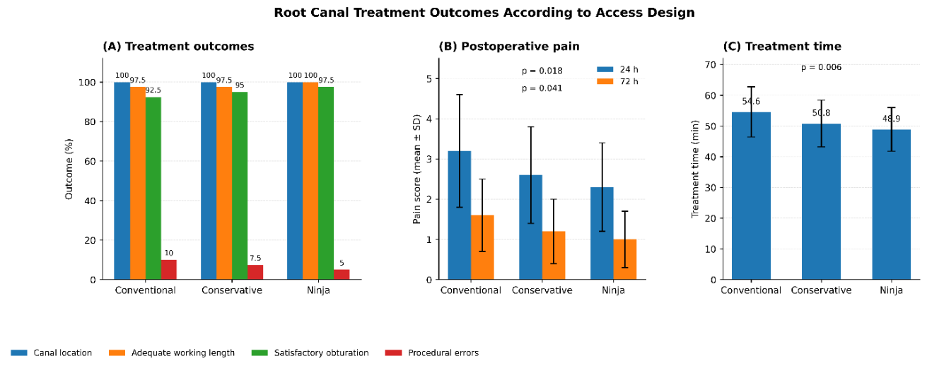
Figure 1: Root canal treatment outcomes according to access design. (A) Canal location, adequate working length, satisfactory obturation, and procedural errors expressed as percentages; (B) postoperative pain scores at 24 and 72 hours presented as mean ± SD; and (C) treatment time presented as mean ± SD. p-values indicate overall comparisons among the conventional, conservative, and Ninja access groups.
Table IV presents the multivariable predictors of fracture resistance. Conservative access (β=0.31; 95% CI, 0.12–0.51; p=0.002), Ninja access (β=0.46; 95% CI, 0.25–0.67; p<0.001), and greater remaining dentin thickness (β=0.52; 95% CI, 0.34–0.70; p<0.001) were positively associated with fracture resistance. Canal curvature (β=−0.18; 95% CI, −0.31 to −0.05; p=0.008) and treatment time (β=−0.14; 95% CI, −0.27 to −0.01; p=0.036) showed negative associations, while patient age was not significant (β=−0.09; 95% CI, −0.21 to 0.03; p=0.142).
Table IV: Multivariable Predictors of Fracture Resistance and Successful Root Canal Treatment(n=120)
| Predictor |
β / OR |
95% CI |
p-value |
| Conservative access design |
0.31 |
0.12–0.51 |
0.002 |
| Ninja access design |
0.46 |
0.25–0.67 |
<0.001 |
| Remaining dentin thickness |
0.52 |
0.34–0.70 |
<0.001 |
| Canal curvature |
−0.18 |
−0.31 to −0.05 |
0.008 |
| Patient age |
−0.09 |
−0.21 to 0.03 |
0.142 |
| Treatment time |
−0.14 |
−0.27 to −0.01 |
0.036 |
Note: β values represent standardized regression coefficients for fracture resistance. Higher remaining dentin thickness and minimally invasive access designs were associated with greater fracture resistance. Statistical significance was defined as p<0.05.
The present study demonstrated that minimally invasive access cavity designs were associated with greater fracture resistance than conventional access preparation, while major root canal treatment outcomes remained comparable among the groups. These findings suggest that conservative access preparation may preserve structural integrity without substantially compromising key technical aspects of root canal treatment.
DISCUSSION
The present study found that minimally invasive endodontic access designs were associated with higher fracture resistance than conventional access, with the Ninja access group showing the greatest mean resistance. Conservative and Ninja access also maintained comparable canal location, working-length achievement, obturation quality, and procedural-error rates. These findings support the principle that preservation of coronal and pericervical dentin may improve the mechanical behavior of root-filled teeth. Recent systematic reviews similarly report greater load capacity or fracture resistance with conservative access compared with traditional preparation, although substantial heterogeneity exists among studies 14.
The superiority of the Ninja and conservative groups may be explained primarily by greater preservation of dentin rather than by the access configuration itself. The current regression analysis supports this interpretation because remaining dentin thickness was the strongest positive predictor of fracture resistance. A study showed that contracted access preserved significantly more coronal and pericervical dentin while maintaining comparable instrumentation outcomes, whereas some other study found superior fracture resistance for conservative and truss designs after thermocycling and dynamic loading 15,16. Conversely, evidence is not completely consistent; it is reported no fracture-resistance difference among traditional, conservative, and truss access, and similarly, no advantage of ultraconservative access in mandibular molars was found 17,18. Differences in tooth morphology, restorative procedures, instrumentation systems, loading conditions, and the amount of structural loss probably account for these discrepancies.
An important finding was that the improved fracture resistance in the minimally invasive groups was not accompanied by deterioration in the principal technical outcomes of root canal treatment. Canal location and working-length achievement were essentially equivalent across groups, while satisfactory obturation was numerically more frequent with conservative and Ninja access. This observation is clinically relevant because restricted access has traditionally been criticized for compromising visibility, negotiation, instrumentation, and filling 19. Nevertheless, systematic evidence remains cautious, as it was found that most studies assessing contracted cavities reported some reduction in instrumentation efficacy 20. Whereas a study concluded that minimally invasive preparations may create potential concerns regarding disinfection, filling, and procedural errors 21. The lower postoperative pain scores at 24 and 72 hours and the shorter treatment time observed with minimally invasive designs provide additional clinical support for their use in appropriately selected cases. However, the pain findings should be interpreted cautiously because postoperative discomfort is multifactorial and may depend on pulpal/periapical status, instrumentation, irrigation, occlusal adjustment, and analgesic use. Importantly, a randomized trial found no significant difference in postoperative pain between conservative and truss access despite differences in bacterial reduction, suggesting that access design alone may not reliably determine postoperative symptoms 22.
The current study is limited by the relatively modest sample size, short postoperative follow-up, restriction to single-rooted teeth, and use of laboratory fracture testing, which does not reproduce complex long-term intraoral fatigue. In addition, the absence of long-term radiographic healing and tooth-survival outcomes limits conclusions about overall endodontic success. Moreover, laboratory fracture resistance is a surrogate mechanical outcome and does not directly represent long-term clinical tooth survival. Future multicenter randomized studies should evaluate diverse tooth types, longer follow-up, three-dimensional imaging, cyclic loading, microbial reduction, and restoration-related variables to determine whether the structural advantage of minimally invasive and Ninja access translates into superior long-term clinical survival.
CONCLUSION
The access design with the least amount of access was associated with the highest fracture resistance, with Ninja access having the highest fracture resistance. In both cases, the canal localization, working length achievement and obturation quality remained almost equal, but in the conservative group was associated with less postoperative pain and the treatment time was shorter. A preservation of the coronal and pericervical dentin may increase the mechanical resistance of the root-filled teeth. A long-term clinical survival, however, was not evaluated. Further studies should consider the survival of teeth, periapical healing and function of teeth with various tooth types.
References
- El-Ma'aita A, Qualtrough A, Darcey J, Hunter MJ. Rethinking root canal treatment: understanding the difference between preventing and treating endodontic infection. Br Dent J. 2015;219(1):13-16. https://doi.org/10.1038/sj.bdj.2015.527
- Batra D, Dixit A, Marwaha J, Maiti N, Gowda T, Saha A. Effect of obturation techniques on fracture resistance of endodontically treated teeth. Bioinformation. 2025;21(3):361-364. https://doi.org/10.6026/973206300210361
- Abdellatif D, Capar ID, De Fontaine S, Iandolo A, Meyer C, Mancino D. Access cavity in endodontics: balancing precision, preservation, and clinical needs. J Conserv Dent Endod. 2025;28(6):573-587. https://doi.org/10.4103/JCDE.JCDE_270_25
- Akıncı L, Mungan NS. Endodontic access cavities: current approaches to design principles and clinical outcomes. Cumhuriyet Dent J. 2025;28(3):452-461. https://doi.org/10.7126/cumudj.1667325
- Chan MYC, Cheung V, Lee AHC, Zhang C. A literature review of minimally invasive endodontic access cavities - past, present and future. Eur Endod J. 2022;7(1):1-10. https://doi.org/10.14744/eej.2022.62681
- Rane S, Pandit V, Gaikwad A, Chavan S, Patil R, Shinde M. Minimally invasive access cavity designs: a review. J Pharm Bioallied Sci. 2024;16(Suppl 3):S1971-S1973. https://doi.org/10.4103/jpbs.jpbs_450_24
- Haralur SB, Al-Qahtani AS, Al-Qarni MM, Al-Homrany RM, Aboalkhair AE. Influence of remaining dentin wall thickness on the fracture strength of endodontically treated tooth. J Conserv Dent. 2016;19(1):63-67. https://doi.org/10.4103/0972-0707.173201
- Sevin M, Orio E, Collignon A-M. Minimally invasive access cavities: a benefit/risk analysis. J Clin Med. 2025;14(7):2476. https://doi.org/10.3390/jcm14072476
- Kwon SK, Seog M, Kim D, Jung IY. Impact of minimally invasive root canal treatment on healing outcomes in a randomized clinical trial. Sci Rep. 2025;15(1)):23260. https://doi.org/10.1038/s41598-025-02905-z
- Chaudhary S, Mishra P, Singh SK, Verma P, Khan R, Upadhayay A. Comparative evaluation of fracture resistance in endodontically treated teeth: an analysis of different restorative materials. J Pharm Bioallied Sci. 2025;17(Suppl 2):S1650-S1652. https://doi.org/10.4103/jpbs.jpbs_216_25
- Mrinalini M, Gupta A, Soi S, Abraham D, Bukhari SH. Endodontic access cavity design and fracture resistance: a systematic review and meta-analysis of conventional vs. newer access cavity. Cureus. 2024;16(9):e68796. https://doi.org/10.7759/cureus.68796
- Makati D, Shah NC, Brave D, Rathore VPS, Bhadra D, Dedania MS. Evaluation of remaining dentin thickness and fracture resistance of conventional and conservative access and biomechanical preparation in molars using cone-beam computed tomography: an in vitro study. J Conserv Dent. 2018;21(3):324-327. https://doi.org/10.4103/JCD.JCD_311_17
- Sullivan KM, Dean A, Soe MM. OpenEpi: a web-based epidemiologic and statistical calculator for public health. Public Health Rep. 2009;124(3):471–474. https://doi.org/10.1177/003335490912400320
- Ballester B, Giraud T, Ahmed HM, Nabhan MS, Bukiet F, Guivarc’h M. Current strategies for conservative endodontic access cavity preparation techniques—systematic review, meta-analysis, and decision-making protocol. Clin Oral Invest. 2021;25(11):6027-44. https://doi.org/10.1007/s00784-021-04080-7
- Peng W, Zhou X, Gao Y, Xu X. Effect of access cavity preparation on dentin preservation, biomechanical property, and instrumentation efficacy: a micro-computed tomographic study. J Endod. 2022;48(5):659-668. https://doi.org/10.1016/j.joen.2021.12.012
- Santosh SS, Ballal S, Natanasabapathy V. Influence of minimally invasive access cavity designs on the fracture resistance of endodontically treated mandibular molars subjected to thermocycling and dynamic loading. J Endod. 2021;47(9):1496-1500. https://doi.org/10.1016/j.joen.2021.06.020
- Barbosa AF, Silva EJ, Coelho BP, Ferreira CM, Lima CO, Sassone LM. The influence of endodontic access cavity design on the efficacy of canal instrumentation, microbial reduction, root canal filling and fracture resistance in mandibular molars. Int Endod J. 2020;53(12):1666-1679. https://doi.org/10.1111/iej.13383
- Augusto CM, Barbosa AF, Guimarães CC, Lima CO, Ferreira CM, Sassone LM, et al. A laboratory study of the impact of ultraconservative access cavities and minimal root canal tapers on the ability to shape canals in extracted mandibular molars and their fracture resistance. Int Endod J. 2020;53(11):1516-1529. https://doi.org/10.1111/iej.13369
- Özyürek T, Ülker Ö, Demiryürek EÖ, Yılmaz F. The effects of endodontic access cavity preparation design on the fracture strength of endodontically treated teeth: traditional versus conservative preparation. J Endod. 2018;44(5):800-5. https://doi.org/10.1016/j.joen.2018.01.020
- Shroff M, Kishan KV, Shah N, Saklecha P. Impact of contracted endodontic cavities on instrumentation efficacy—a systematic review. Aust Endod J. 2023;49(1):202-212. https://doi.org/10.1111/aej.12679
- Shabbir J, Zehra T, Najmi N, Hasan A, Naz M, Piasecki L, et al. Access cavity preparations: classification and literature review of traditional and minimally invasive endodontic access cavity designs. J Endod. 2021;47(8):1229-1244. https://doi.org/10.1016/j.joen.2021.05.007
- Özen MM, Karataş E. Effect of cavity design on bacterial reduction in root canals and postoperative pain level: a randomised controlled trial. Aust Endod J. 2025;51(3):632-639. https://doi.org/10.1111/aej.12976

Journal of Dentistry and Oral Sciences Research, J Dent Oral Sci Res 2026:1(1), p15-19 (jdosr.org)
© 2026 Authors. This work is published by Multidisciplinary Scholarly Advancement and Research MSAR Institute. The full terms of Journal Publishing policy is available at https://jdosr.org/index.php/jdosr/publishing-policy and incorporate the Creative Commons Attribution – Non Commercial (CC BY, NC 4.0) License https://creativecommons.org/licenses/by-nc/4.0/. By accessing the work you hereby accept the Terms. Non-commercial uses of the work are permitted without any further permission, provided the work is properly attributed. Publisher’s Note: MSAR Institute remains neutral with regard to jurisdictional claims in published maps and institutional affiliations, and assumes no liability for the scientific accuracy or clinical efficacy of the content herein, as they rest entirely with the authors.