
Comparative Effect of Minimally Invasive Endodontic Access Cavity Designs on Fracture Resistance and Root Canal Treatment Outcomes
DOI:
https://doi.org/10.68041/jdosr.v1i1/04Keywords:
Dentin, Endodontics, Postoperative Pain, Root Canal Obturation, Root Canal Preparation, Tooth FracturesAbstract
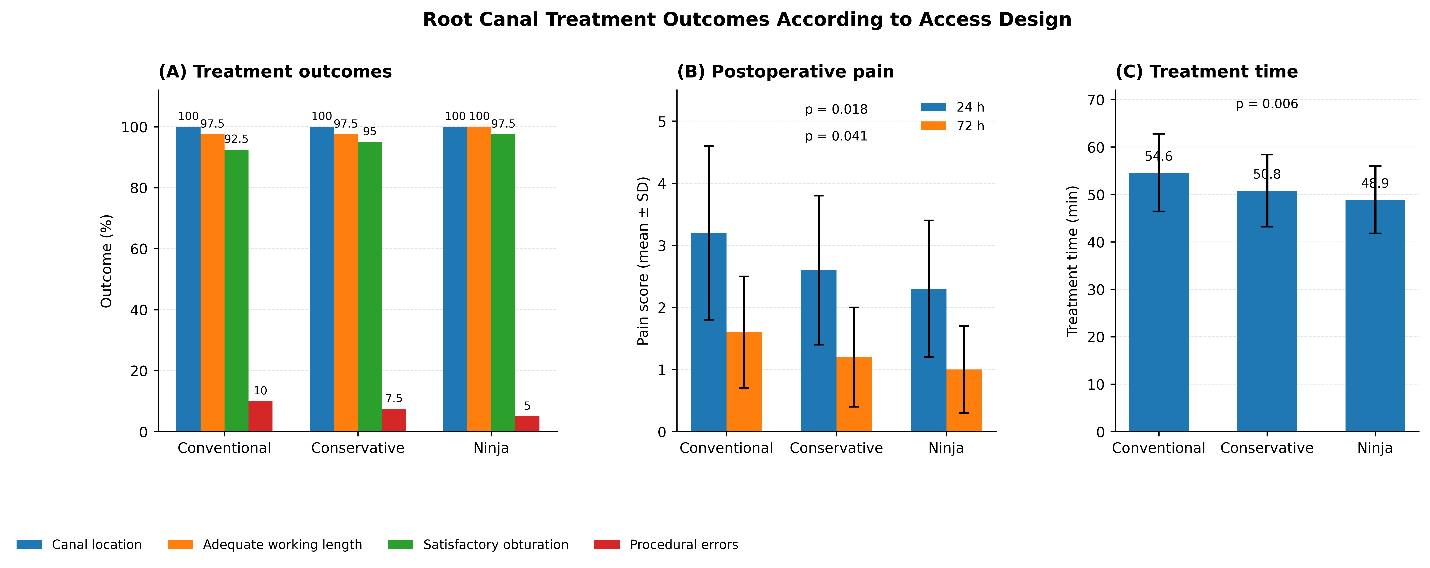
Background: Minimally invasive designs seek to maintain pericervical dentin and can improve the resistance of root-filled teeth. This study aimed to compare the fracture resistance and outcomes of conventional, conservative and ninja endodontic access cavity designs. Methods: 120 single-rooted teeth were allocated to three pre-defined groups (n=40 each): conventional, conservative, and ninja endodontic access cavity. Standardized access preparation, instrumentation, irrigation, and obturation were carried out. The fracture resistance was tested on a testing machine. Canal location, the working length achieved, quality of obturation, procedural errors, postoperative pain, treatment time, canal curvature, and remaining dentin thickness were evaluated. All data were analyzed using SPSS version 26.0, where p<0.05 was considered statistically significant. Results: There was a significant difference in fracture resistance between the groups (p<0.001), with the lowest resistance in the conventional access group (742.6 ± 118.4 N), followed by the conservative access group (861.3 ± 126.7 N) and the ninja endodontic access cavity (918.7 ± 134.2 N). Canal location, working length, obturation quality, and procedural errors were not significantly different. Postoperative pain was lower at 24 hours (p=0.018) and 72 hours (p=0.041) in the minimally invasive groups, and the duration of treatment was also shorter (p=0.006). The remaining dentin thickness was positively associated with fracture resistance (p<0.001), while canal curvature had a negative association (p=0.008). Conclusion: Conservative and ninja endodontic access cavities were associated with better fracture resistance without compromising treatment outcomes. Longer-term studies are needed to determine their effects on tooth survival and periapical healing.
References
1. El-Ma'aita A, Qualtrough A, Darcey J, Hunter MJ. Rethinking root canal treatment: understanding the difference between preventing and treating endodontic infection. Br Dent J. 2015;219(1):13-16. https://doi.org/10.1038/sj.bdj.2015.527
2. Batra D, Dixit A, Marwaha J, Maiti N, Gowda T, Saha A. Effect of obturation techniques on fracture resistance of endodontically treated teeth. Bioinformation. 2025;21(3):361-364. https://doi.org/10.6026/973206300210361
3. Abdellatif D, Capar ID, De Fontaine S, Iandolo A, Meyer C, Mancino D. Access cavity in endodontics: balancing precision, preservation, and clinical needs. J Conserv Dent Endod. 2025;28(6):573-587. https://doi.org/10.4103/JCDE.JCDE_270_25
4. Akıncı L, Mungan NS. Endodontic access cavities: current approaches to design principles and clinical outcomes. Cumhuriyet Dent J. 2025;28(3):452-461. https://doi.org/10.7126/cumudj.1667325
5. Chan MYC, Cheung V, Lee AHC, Zhang C. A literature review of minimally invasive endodontic access cavities - past, present and future. Eur Endod J. 2022;7(1):1-10. https://doi.org/10.14744/eej.2022.62681
6. Rane S, Pandit V, Gaikwad A, Chavan S, Patil R, Shinde M. Minimally invasive access cavity designs: a review. J Pharm Bioallied Sci. 2024;16(Suppl 3):S1971-S1973. https://doi.org/10.4103/jpbs.jpbs_450_24
7. Haralur SB, Al-Qahtani AS, Al-Qarni MM, Al-Homrany RM, Aboalkhair AE. Influence of remaining dentin wall thickness on the fracture strength of endodontically treated tooth. J Conserv Dent. 2016;19(1):63-67. https://doi.org/10.4103/0972-0707.173201
8. Sevin M, Orio E, Collignon A-M. Minimally invasive access cavities: a benefit/risk analysis. J Clin Med. 2025;14(7):2476. https://doi.org/10.3390/jcm14072476
9. Kwon SK, Seog M, Kim D, Jung IY. Impact of minimally invasive root canal treatment on healing outcomes in a randomized clinical trial. Sci Rep. 2025;15(1)):23260. https://doi.org/10.1038/s41598-025-02905-z
10. Chaudhary S, Mishra P, Singh SK, Verma P, Khan R, Upadhayay A. Comparative evaluation of fracture resistance in endodontically treated teeth: an analysis of different restorative materials. J Pharm Bioallied Sci. 2025;17(Suppl 2):S1650-S1652. https://doi.org/10.4103/jpbs.jpbs_216_25
11. Mrinalini M, Gupta A, Soi S, Abraham D, Bukhari SH. Endodontic access cavity design and fracture resistance: a systematic review and meta-analysis of conventional vs. newer access cavity. Cureus. 2024;16(9):e68796. https://doi.org/10.7759/cureus.68796
12. Makati D, Shah NC, Brave D, Rathore VPS, Bhadra D, Dedania MS. Evaluation of remaining dentin thickness and fracture resistance of conventional and conservative access and biomechanical preparation in molars using cone-beam computed tomography: an in vitro study. J Conserv Dent. 2018;21(3):324-327. https://doi.org/10.4103/JCD.JCD_311_17
13. Sullivan KM, Dean A, Soe MM. OpenEpi: a web-based epidemiologic and statistical calculator for public health. Public Health Rep. 2009;124(3):471–474. https://doi.org/10.1177/003335490912400320
14. Ballester B, Giraud T, Ahmed HM, Nabhan MS, Bukiet F, Guivarc’h M. Current strategies for conservative endodontic access cavity preparation techniques—systematic review, meta-analysis, and decision-making protocol. Clin Oral Invest. 2021;25(11):6027-44. https://doi.org/10.1007/s00784-021-04080-7
15. Peng W, Zhou X, Gao Y, Xu X. Effect of access cavity preparation on dentin preservation, biomechanical property, and instrumentation efficacy: a micro-computed tomographic study. J Endod. 2022;48(5):659-668. https://doi.org/10.1016/j.joen.2021.12.012
16. Santosh SS, Ballal S, Natanasabapathy V. Influence of minimally invasive access cavity designs on the fracture resistance of endodontically treated mandibular molars subjected to thermocycling and dynamic loading. J Endod. 2021;47(9):1496-1500. https://doi.org/10.1016/j.joen.2021.06.020
17. Barbosa AF, Silva EJ, Coelho BP, Ferreira CM, Lima CO, Sassone LM. The influence of endodontic access cavity design on the efficacy of canal instrumentation, microbial reduction, root canal filling and fracture resistance in mandibular molars. Int Endod J. 2020;53(12):1666-1679. https://doi.org/10.1111/iej.13383
18. Augusto CM, Barbosa AF, Guimarães CC, Lima CO, Ferreira CM, Sassone LM, et al. A laboratory study of the impact of ultraconservative access cavities and minimal root canal tapers on the ability to shape canals in extracted mandibular molars and their fracture resistance. Int Endod J. 2020;53(11):1516-1529. https://doi.org/10.1111/iej.13369
19. Özyürek T, Ülker Ö, Demiryürek EÖ, Yılmaz F. The effects of endodontic access cavity preparation design on the fracture strength of endodontically treated teeth: traditional versus conservative preparation. J Endod. 2018;44(5):800-5. https://doi.org/10.1016/j.joen.2018.01.020
20. Shroff M, Kishan KV, Shah N, Saklecha P. Impact of contracted endodontic cavities on instrumentation efficacy—a systematic review. Aust Endod J. 2023;49(1):202-212. https://doi.org/10.1111/aej.12679
21. Shabbir J, Zehra T, Najmi N, Hasan A, Naz M, Piasecki L, et al. Access cavity preparations: classification and literature review of traditional and minimally invasive endodontic access cavity designs. J Endod. 2021;47(8):1229-1244. https://doi.org/10.1016/j.joen.2021.05.007
22. Özen MM, Karataş E. Effect of cavity design on bacterial reduction in root canals and postoperative pain level: a randomised controlled trial. Aust Endod J. 2025;51(3):632-639. https://doi.org/10.1111/aej.12976
Published
Issue
Section
License
Copyright (c) 2026 Naila Anjum, Inam Ur Rehman, Faiza Ikram

This work is licensed under a Creative Commons Attribution-NonCommercial 4.0 International License.
Journal of Dentistry and Oral Sciences Research (JDOSR) follows the Attribution Creative Commons-Non commercial (CC BY-NC 4.0) license which allows the users to copy and redistribute the material in any medium or format, remix, transform and build upon the material. The users must give credit to the source and indicate, provide a link to the license, and indicate if changes were made. However, the CC BY-NC 4.0 license restricts the use of material for commercial purposes (Further details are available at website. The Editorial Board of the JDOSR endeavors to ensure the accuracy, integrity, and quality of all published content. However, the responsibility of opinions, interpretations, and conclusions expressed in the published articles rests entirely with the authors and do not necessarily reflect the views of the Editorial Board, publisher, or affiliated institutions.